MRI
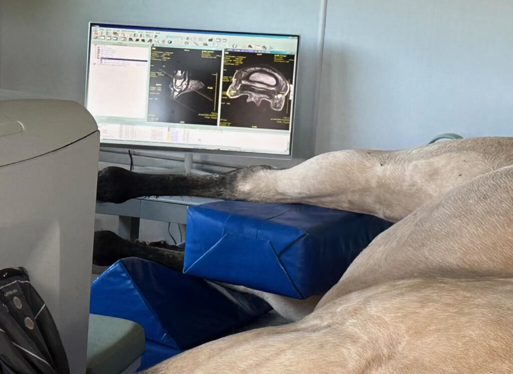
We are excited to offer Montana's only equine Magnetic Resonance Imaging (MRI)Magnetic resonance imaging (MRI) is a painless and non-invasive imaging tool. First used in human medicine in the 1980s, MRI imaging relies on a strong magnet to create detailed medical images. The MRI’s magnetic field is thousands of times more powerful than the earth’s own magnetic field, and briefly aligns the “spin” of atoms in the area being imaged. A computer then converts the resulting radiofrequency energy into detailed three-dimensional images of the tissues in the patient’s body.
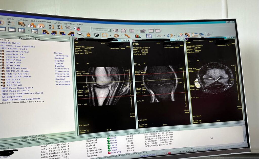
Since about 2010, MRI has been increasing used in equine veterinary medicine, revolutionizing veterinarians’ ability to diagnose and treat problems that cannot be evaluated with conventional tools like radiographs or ultrasound. This tool is primarily used to diagnose orthopedic issues in the lower limb. Without MRI, it is difficult or impossible to see many critical structures, especially soft tissues like tendons, ligaments, and cartilage in a horse’s foot. As it turns-out, these structures are among the most frequent contributors to chronic discomfort and associated performance issues. In particular, MRI is usually strongly indicated for horses with chronic discomfort localized to the foot or other area in the lower limbs where ultrasound and radiographs are inconclusive
That means that MRI can play a critical role in helping us diagnose and treat specific issues that might otherwise be reliant upon an educated guess or a “shot-in-the-dark”, or otherwise untreatable. With the addition of this modality in the Summer of 2023, Montana Equine is proud to be playing a leading role in helping to advance the science of equine imaging in our region.
What can MRI Identify?
Typical problems that MRI can identify even when radiographs or ultrasound do not confirm significant abnormalities:
- Bone and Joint Inflammation
- Synovitis
- Cartilage Defects
- Sub-chondral and Physeal Lesions
- Ligament Inflammation
- Suspensory Desmitis
- Check Ligament Desmitis
- Impar Ligament Desmitis
- Sesamoidean Ligament Desmitis
- Collateral Ligament Desmitis (especially of the coffin joint)
- “Navicular Issues” aka “Caudal Heel Syndrome”
- Bone Edema
- Navicular Bursitis
- Deep Flexor Tendonitis (inside the foot)
- Tendon Issues
- Deep Digital Flexor Tendonitis (especially when within the foot)
Have Questions?
Please feel free to call Dr. Peter Heidmann, DVM DACVIM or Shawni Hansen CVT at 406-285-0123 if you would like to learn more about how we use MRI in clinical practice.
FOR VETERINARIANS:
FOR VETERINARIANS:
If you’d like to discuss referral of your clients and patients for MRI imaging, please feel free to call Dr. Peter Heidmann, Shawni Hansen, or any of the attending veterinarians at Montana Equine.
If you’ve already determined that you would like to refer your clients or patients for MRI, please complete this form to expedite referral for imaging.